A
NOVEL INSTRUMENT FOR SURFACE ANALYSIS
PEEM is an electron microscope imaging technique. A surface is illuminated by ultraviolet (UV) radiation. The UV light generates photoelectrons for which intensity greatly depends on the chemical composition and the topography of the surface. The photoelectrons are accelerated into the microscope column, pass through a combination of magnifying lenses, and are projected onto a detector, converting the electron image into a visible image. The visible image displays the strongly magnified distribution of photoelectrons emitted from the surface. This image can be recorded at video rate using a CCD camera, allowing fast changes occurring on the surface to be recorded. The magnification can be easily controlled by voltages applied to the electrostatic lenses and can be electronically adjusted in a range up to
x8000.
Besides this imaging capability, the improved design of the STAIB-PEEM offers the possibility of measuring the energy distribution of photoelectrons to characterize the atomic states on the sample. Energy analyses are possible on the complete image at full video speed, or on a small size spot at higher sensitivity and larger dynamic range. The Imaging Energy Filter (IEF) can be mounted in-line between the detector and the microscope column. The Micro Spot Analysis Filter (MAF) is mounted on the side of the column. A stigmatic beam switch allows instant change between analysis modes while preserving the image
quality. 
PEEM-350: A STATE OF THE ART INSTRUMENT FOR SURFACE
RESEARCH
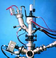
| As a microscope technique, PEEM can be used in a wide range of applications. Because each investigation requires a slightly different technical configuration, the STAIB-PEEM-350 is designed as a flexible, modular system. The instrument offers maximum capabilities for microscopy, energy filtering, and microanalysis, while remaining very compact and extremely stable mechanically. For instance, the detector part, a large chevron channel plate with fiber optic coupling, is fully integrated into the optical column, allowing highest spatial resolution capability even with the PEEM mounted in complex environmental conditions.
Should vibration of the whole system be a limiting factor for the resolution, an integrated sample holder can be
incorporated.
|
|
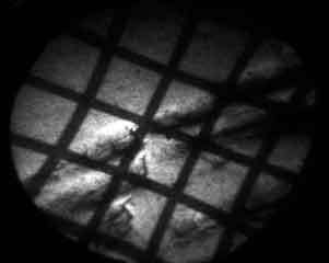
Direct image obtained with a Mercury UV lamp
|
|
|
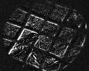
Energy filtered image using the IEF
|
|
The stigmatic beam switch unit is also fully integrated for maximum stability. The stigmatic beam switch provides an accurate adjustment of the selected microscopic area. Only electronic switching, without mechanical adjustment, is needed. The optical column is precisely assembled so that no further time consuming mechanical adjustment of the diaphragm is required. All fine adjustments of the system are controlled by the electronics, keeping set up time to a minimum, even
for inexperienced users. Furthermore, the PEEM Computer Control and Data Acquisition system
automates all major operations in a user friendly manner, ideally suited for operation by different operators. |
The stable and compact design of the PEEM
Microanalyzer allows many custom specific modifications for special, more
sophisticated, investigations. The STAIB INSTRUMENTS team technically supports
these modifications. Examples of customized implementations of the basic
PEEM-350 design include implementing coaxial ion beam excitation (patented
design), adding special detectors for time-of-flight analyses, incorporating a
spin sensitive detector, building a special sample holder for in-situ device
testing.
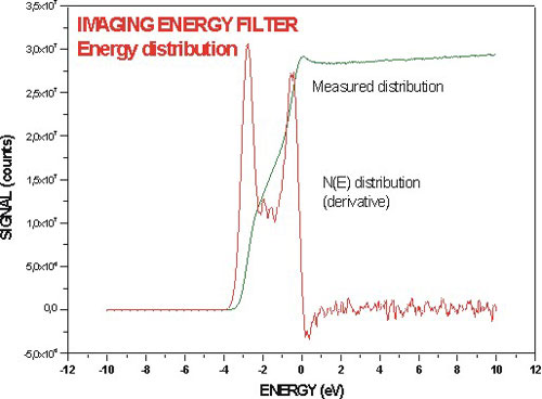
Energy distribution measured with the IEF
ONE
INSTRUMENT, MULTIPLE ILLUMINATION CAPABILITIES
The compact PEEM-350 system can be mounted and combined with many different sources of illumination. The most common sources are:
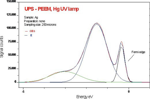
- UV lamps (mercury short arc or deuterium discharge) with proper UV optical benches. With a photon energy of approximately 4.9 eV, UV lamps provide maximum chemical contrast and highest spatial
resolution. Energy analysis reveals the detailed structure of the band near the fermi level and allows maximal chemical
contrast.
- Vacuum UV sources, such as the Helium discharge lamp emitting at 21.2 eV, are used to investigate deeper energy states and allow a
direct comparison with data known from standard UPS techniques. Using the
imaging energy filter, the chemical contrast is greatly enhanced, and the
distribution of elements can be displayed. With the microspotanalyzer, detailed
energy distributions are
obtained.
- X-ray imaging is achieved using
a low energy X-ray source. This technique allows the measurement of XPS distributions
with excellent spatial resolution and with sensitivity limited only by
the intensity of the X-ray source.
- Synchrotron Radiation offers a wide
range of tunable energies at very high intensities. This preferred mode of excitation at higher photon energies requires a more complex installation and beam access to a synchrotron machine. The high intensity and brightness of the illumination allow the measurement of photoelectron spectra at higher kinetic
energies.
- Electron beam excitation provides a different contrast capability. When used
simultaneously with a UV source, charging effects can be reduced. Use of a fine focused electron beam allows for scanning electron microscopy (SEM).
- Ion beam excitation, in low eV to MeV energy range, leads to exciting new imaging techniques.
ONE INSTRUMENT, MULTIPLE ANALYSIS
CAPABILITIES
The STAIB-PEEM-350 is a true microanalyzer in the sense that the beam paths for imaging and for spectroscopy are well
separated. This separation enables sharp, high magnification imaging and high resolution energy filtering, which require different beam adjustments. The use of a beam switch inside the microscope column not only makes the design very compact and stable, but also offers a precise transition between imaging and analysis modes. As the beam switch is a stigmatic device, the image quality is preserved up to the entrance of the
Microspot Analysis Filter (MAF). The entrance diaphragm picks up a well defined sample area, commonly adjusted in the micrometer
range. The diaphragm is fixed in position requiring no time consuming mechanical
adjustments. The image can be scanned over the diaphragm and an image corresponding to a well defined photoelectron energy can be measured. In addition, the
Imaging Energy Filter (IEF) maintains both fast imaging capability and energy filtering. A newly designed optical system accurately filters a complete PEEM image over the full detector area. The unfiltered image is resolved into single distributions of different energies. Each distribution can be assigned to a specific work function, i.e., to a specific chemical phase of the sample. The new design results in energy filtering of the complete image and fast acquisition, a unique feature for the observation of time dependent systems.
STAIB-PEEM, A DESIGN WITH PROVEN PERFORMANCES FOR MANY
APPLICATIONS
Ten years after its first commercialization, the STAIB PEEM has grown as the commercial PEEM system used by the largest community of users with very different applications. Constant product care and R&D efforts are essential to fulfill users requirements. STAIB INSTRUMENTS is proud that, based on PEEM results, major technical improvements, discoveries, and patented inventions have been
made by our users. The following is a partial list of these applications:
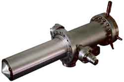
PEEM 350 equipped with the Imaging Energy Filter and
the Microspot Analysis Filter |
- Semiconductor wafer analysis and chemical control, using chemical imaging and video speed imaging
- Superconductor characterization and control of device function
- Improvement of field emission display
technology
- Particle analysis using special preparation techniques
- Imaging of magnetic structures
- In-situ observation of catalytic reactions
for environmental studies using differential pumping
- Time-of-flight ion imaging spectroscopy
- In-situ control of growth
of specific crystal phases
- Characterization of organic films
- Study of multiple photon ionization by
laser excitation.
|
This research is conducted in major national and industrial
laboratories worldwide. In order to preserve the investment value of our equipment
and to benefit from our new developments STAIB offers an upgrade guaranty for
newly developed systems: there is no obsolescence for STAIB
INSTRUMENTS products.
Ask about PEEM-FACTS for more
information.
PEEM-350
HIGHLIGHTS
- Unique compact and high stability design, as a stand alone instrument or as a component
- Mounting in any position, with optional retracting device
- Very large field of view using a of 40 mm diameter fiber optic detector
- Magnification up to x8000
- High spatial resolution 40 nm on chemical contrast, better using topographical contrast
- Energy filtering of complete images at any magnification and at video
rate
- Energy filtering of selected micro size areas with electrical imaging capability
- Fully UHV compatible, bakeable to 250°C
- Factory aligned column electronically tunable (no mechanical adjustment)
- Fits standard CF flanges, ideally suited as an add-on instrument on any complex
systems
- Differential pumping capability for working at higher pressure
- Computer controls for the microscope, CCD camera, and Electron Spectroscopy
- Multi-tasking software capability for PCs with simultaneous image and spectrum acquisition
capability.
Ask about PEEM-SPECS for more information.
PEEM FAMILY OF
UPGRADABLE SYSTEMS
Basic configuration with detector
- larger magnification
- stigmator
- Imaging Energy Filter (IEF)
- Microspot Analysis Filter (MAF)
- combined IEF and MAF
PEEM VACUUM CHAMBERS
AND COMPLETE UHV SYSTEMS
STAIB INSTRUMENTS offers stand alone complete systems
including multiple deposition and
analysis technique, for example combining STM, SEM, PEEM, LEED, AES, XPS, or Ellipsometry with ion etching and deposition sources.
STAIB INSTRUMENTS offers a functional guaranty and technical postsales support
worldwide.
- Compact UHV chamber for PEEM with accessories
- Multitechnique UHV system for PEEM combined other
techniques
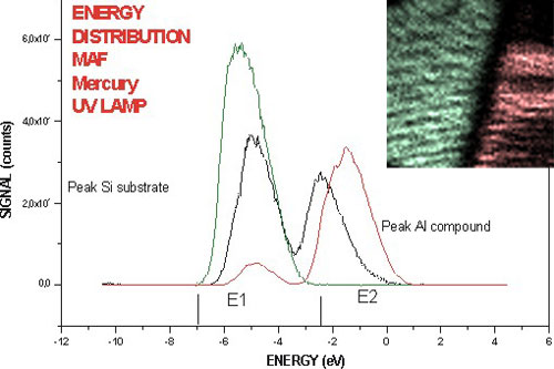
Energy distribution and scanning image obtained with MAF
Ask about PEEM-SYSTEMS for more
information.
PEEM
ACCESSORIES
- Differential pumping option
- Mechanical retraction stage
- Mercury Discharge UV lamp and optic
Deuterium Discharge UV lamp and optic
- Vacuum UV Helium Discharge lamp
- Integrated sample holder
- Standard sample holder with heating, cooling
capability
- PEEM
Control and PEEM
Vision
- Ion gun for sample cleaning
- Dual anode X-ray source
- Electron gun for sample Illumination and Scanning Electron
Microscopy.
Energy distribution after ion cleaning and deconvolution
Contact our sales department for actual information.
  
|